|
Impacted
wisdom tooth
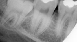
The following
pictures, were taken for surgical extraction of lower 3rd molar
for 21 years old female who attend oral surgery clinic in
Alkarama specialized dentistry center/Baghdad. These pictures
can be used as a guide for beginner dentists, as it illustrates,
step by step, the procedure for removal of some how straight
forward case of class 1, b impacted lower 3rd molar (right
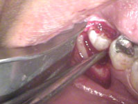
side), Picture no 1
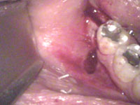
Picture no 2
shows the shape of flap after incision making, which is mostly 2
sided flap with vertical incision extends from distal aspect of
lower 1st molar slightly above the buccal vestibule, upward and
backward involving distal gingival of lower 2nd molar tooth. The
horizontal incision extends in the mid way between buccal and
lingual sides to the origin of external oblique ridge.
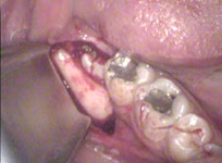
Picture no 3
shows the surgical field after flap retraction. Notice how it
should expose sufficient bone mesial, distal and downward
directions.
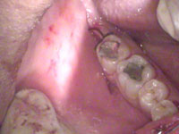
Picture no 4
shows the surgical field after bone removal which should expose
depth of the root by creating a tunnel like opening with
additional bone removal from mesial aspect of the tooth for
elevator application as in picture no 5
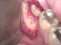
no 4
no 5
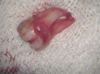
Picture no 6
shows the final step in this procedure. As the picture shows one
stich may be enough to close the surgical wound.
It's mandatory to irrigate e the wound by physiological saline
to ensure clean wound post operatively.
This patient was
prophylactic antibiotic cover as single dose preoperatively.
رجوع
|